History of the Cell, Part II
Beyond the Microscope

Our understanding of the biological cell has come a long way since Robert Hooke first saw it under the microscope in 1665, and most of this rapid progress has been in the past 150 years. Before then, cell scientists were just determining that cells are present in all living things and the role of the first known organelle, the nucleus, as explored in the last Biology Bytes article, History of the Cell, Part I.
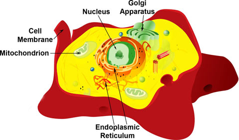
While continuing to better understand the nucleus, researchers also started to investigate other organelles in the cell. Inside each cell are many compartmentalized subunits, each serving a different function in the machinery of the cells; these subunits are called organelles. As vital as the microscope was to first seeing and understanding the cell, many other tools served key roles in elucidating the crucial functions of organelles.
Although the nucleus was known to split in half when cells divide, it was unclear what its exact role was. Only the advent of dyes that selectively stain the nucleus and better methods of preserving samples allowed German biologist Walther Flemming, in 1879, to see tiny “rods” within the nucleus. Due to their visibility, he named them “chromatin” (the prefix “chroma” means “color”). These structures are what we know today as chromosomes, which are tightly packed, organized bundles of DNA within every nucleus: the blueprints for life.
Observing the chromosomes, Flemming studied how the nucleus functions when the cell divides into two new cells. In 1882, he reported that during cell division the chromosomes align in the middle of the nucleus and split lengthwise, becoming equally distributed into the two new cells. However, it was not until the early 1900s that the role of the nucleus in heredity was recognized, after genetics studies using pea plants, done by the Austrian priest Gregor Johann Mendel in the 1850s and 1860s, were rediscovered. Mendel is posthumously known as the “father of genetics.”
We now have a much better understanding of not only how the chromosomes in the nucleus of every cell store a person’s genetic information, and how these genetics are passed on to a new cell or the next generation, but also why, for example, liver cells know to be liver cells and don’t change into brain cells. Although every cell in a body shares the same genome, factors in the nucleus control which genes a given cell uses, controlling the cell’s identity and telling it how to function.
At the same time, researchers started investigating another central feature of the cell: its surrounding membrane. In 1773, the English surgeon William Hewson reported seeing red blood cells shrink and swell depending upon the liquid they were in and proposed that cells were envelope-bound bodies. This is because membranes readily allow water to go inside the cell or leave it (using a process called osmosis). Unfortunately, Hewson’s work was largely unknown and because the cell membrane was difficult to see with the microscopes of the time, there was much debate over whether it existed for some time.
The membrane dilemma was settled at the end of the 1800s by Charles Ernest Overton, at the University of Lund, Sweden. Overton repeated Hewson’s findings, but went further and showed that the membrane is semi-permeable; cells actively select specific molecules to bring inside the cell, and others to transport outside. The membrane was quite real, and more excitingly, it was active! Substantial progress in understanding the membrane was made during the first half of the 1900s. We now understand it has a key role in not only regulating what enters and exits the cell, but also in maintaining the cell structure and communication between cells.
At the turn of the 20th century, all the pieces were in place for researchers to focus on what was going on in the rest of the cell. After the nucleus, one of the next organelles identified was the Golgi apparatus, seen by Italian scientist Camillo Golgi in 1898. As happens often in science, Golgi wasn’t necessarily the first to observe the structure; several other researchers had reported similar structures, but because Golgi was very consistent and clean in his methods and results, his findings were the first widely-accepted. Consistent results were especially difficult because the Golgi apparatus actually varies in shape, making many think it was an artifact of poor microscopes and stains for a long time. But in the 1930s and 40s, researchers established its role in cell secretion. Today, we know the Golgi apparatus is not only involved in secreting large molecules, but also in protein modification. Overall, the Golgi apparatus determines where proteins need to go in the cell, and packages and delivers them, like the cell’s own post office.
By 1940, the study of cells had come as far as it could with just microscopes. Biologists increasingly turned to biochemistry to answer cellular questions. Additionally, around the 1930s a revolutionary investigative tool became available: the ultracentrifuge. The ultracentrifuge (a high-speed centrifuge) can separate particles by size and weight, as it spins samples at very high speeds, up to 1,000,000 times the force of gravity in modern machines.
In 1933, Robert Bensley, at the University of Chicago, used the ultracentrifuge to separate specific organelles from the rest of the cell to better study them. Bensley isolated the mitochondrion (plural mitochondria), which had been recently discovered by German pathologist Richard Altmann. Bridging the gap between seeing the cells under the microscope and understanding their biochemistry, Bensley confirmed his centrifuged product through staining. By the 1950s, researchers were widely applying ultracentrifuge techniques to determine the function of different organelles.
But despite knowing that mitochondria were present, their role in the cell remained murky. This understanding was provided by Albert Claude, at the Rockefeller Institute, who collaborated with biochemists to measure the enzyme activity of the isolated mitochondria. (Enzymes are proteins that help cause specific chemical reactions to take place in the cell.) Mitochondria were found to have important oxidative enzymes, implicating their role in creating energy for the cell. In 1948, Claude accurately described them as “the real power plants of the cell.”
Fascinatingly, mitochondria are also thought to have once been bacteria that became absorbed by the host cell, an idea called the endosymbiotic theory.
Another groundbreaking instrument of the time was the electron microscope. First available in the 1940s, it focuses a beam of electrons the way a normal microscope focuses a ray of light. Today’s electron microscopes can magnify a sample’s image 500,000-fold, but they are limited in the thickness of tissue they can penetrate. In 1945, Claude used an electron microscope to look at mitochondria, but he could not see them clearly as they were too thick. As any professor would do, Claude quickly set the task before the students in his lab.
Keith Porter, a postdoctoral student who joined Claude’s lab in 1939, practically lived under the electron microscope, improving techniques to view cells. In the 1940s, Porter examined a “lace-like reticulum” structure at great lengths, and enhanced ways to image it with the electron microscope. Others were also studying the structure at the time and thought it might be involved in creating proteins. In the 1950s, it was given the name we know it by today: the endoplasmic reticulum. The endoplasmic reticulum is indeed the site where many proteins are made in the cell. It receives its instructions from the nucleus, which it surrounds, and sends the completed proteins to the Golgi apparatus, adjacent to it, for further processing and distribution.
Although not even recognized in the 1940s, by 1970 cell biology was a well-established scientific discipline. The picture of the cell as a functioning machine was coming together: The nucleus directed what proteins needed to be made; the proteins were created in the endoplasmic reticulum and shipped to the Golgi apparatus for distribution throughout the cell; and all the while mitochondria provided the cell its energy. Of course, this is a great simplification; there are many other organelles and processes involved. The cell is so complex that researchers today are discovering new details all the time.
At the University of California, Santa Barbara, several groups research not only the function of cell organelles, but the many molecular communication pathways that are constantly active in every cell.
For more on the early history of cell biology, see William Bechtel’s Discovering Cell Mechanisms: The Creation of Modern Cell Biology, Wikipedia’s “Cell Biology,” and Wikipedia’s “Organelle.”
Biology Bytes author Teisha Rowland is a science writer, blogger at All Things Stem Cell, and graduate student in molecular, cellular, and developmental biology at UCSB, where she studies stem cells. Send any ideas for future columns to her at science@independent.com.